Authors: Benoit Hendrickx, MD, PhD; Karl Waked, MD ; Marc Mespreuve, MD, PhD
Published: May 11th, 2020
Background: The face is known for its extreme variation in vascular anatomy. Furthermore, the rapidly increasing number of filler treatments leads to an increase in severe filler-associated complications (such as skin necrosis and blindness) due to intra-arterial injection. Visualizing a patient’s individual complete facial arterial anatomy in a contrast- and radiation-free way has not been published before. This innovative imaging technique could, therefore, enhance the safety of minimally invasive surgical procedures as it provides a harmless way to map the arteries of the face.
Objectives: Evaluate a newly developed imaging technique to visualize the arteries of the face in a noninvasive and radiation-free manner.
Methods: The individual arterial facial anatomy of 20 volunteers was studied by an imaging technique, combining infrared (IR) facial warming and 3-dimensional (3D) time of flight (TOF) magnetic resonance angiography (MRA). The source and maximum intensity projection images were assessed by 2 investigators, familiar with the anatomy of the face.
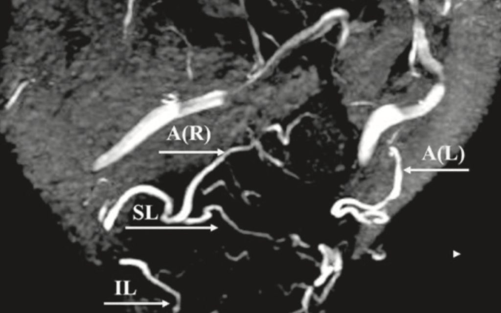
Results: The MRA technique visualized most of the main facial arteries, albeit in a variable way. The main facial branches of the external carotid artery (facial, angular, supralabial, and superficial temporal arteries) were illustrated well, whereas the visualization of the internal carotid branches (supratrochlear and supraorbital arteries) and nasal branches (dorsal nasal and lateral nasal arteries) was less consistent.
Conclusions: The combination of IR “heat-induced enhancement” and a 3D-TOF MRA sequence may actually be an important step toward the visualization of the variable facial vascular anatomy in a noninvasive, radiation-free, and contrast-free manner.
Key takeaways:
- The 3D Time of Flight MOTSA sequence delivers a feasible imaging method to visualize the individual and highly variable arterial anatomy of the patient’s face.
- Pre-heating the face with an infrared lamp before the MRI significantly increases the visualization of all facial vessels, due to the larger caliber and higher visual signal of the arteries.
- Correct patient positioning and scan preparation are crucial to achieve an optimal visualization. More details can be found in the article and in the instructions manual.
- The workflow is completely risk-free for the patients with no complications nor side-effects reported during the study.