Authors: Marc Mespreuve, MD, PhD; Karl Waked, MD ; Barbara Collard, MD; Joris De Ranter, MD; Francis Vanneste, MD; and Benoit Hendrickx MD, PhD
Published: May 11th, 2021
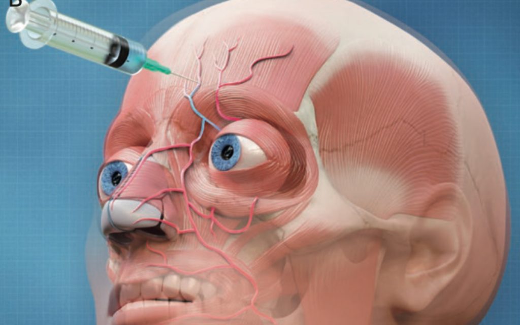
Background: The use of soft tissue fillers for facial rejuvenation is increasing rapidly and the complications, unfortunately, follow the same path. Blindness caused by intravascular filler injections is a rare but devastating complication. Knowledge of the individual arterial anatomy may aid the injector in avoiding injecting into an artery and thus to prevent blindness.
Objectives: To evaluate if the use of magnetic resonance angiography (MRA) may visualize the arterial facial anatomy in a contrast- and radiation-free way and study the individual arterial variations using an augmented reality (AR) image. Methods: The individual arterial anatomy of the 3 terminal branches of the ophthalmic artery (supraorbital [SO]; supratrochlear [STr]; and dorsal nasal [DN] arteries) of 20 volunteers was studied by a 3-Tesla MRI, combining infrared (IR) facial warming and 3-dimensional time-of-flight multiple overlapping thin slab acquisition MRA. The resulting visualization of the facial arteries was shown on the patient’s face through AR technology.
Results: The MRA was able to visualize the SO in 90.0%, STr in 92.5%, and DN arteries in 75% of the examined patients, as well as numerous variations in both vessel localization and path. Furthermore, a proof-of-concept of the AR visualization of the individual arterial anatomy was successfully implemented.
Conclusions: Dermal filler injectors should be aware of the risk of filler-induced blindness and familiarize themselves with the visualization of the variable facial vascular anatomy. The implementation of a one-time MRA and subsequent AR visualization may be useful in the accurate planning of minimally invasive facial rejuvenation procedures.
Key takeaways:
- The new sequence for 3T MRI’s applies a similar workflow as the previously published 1.5T sequence, but results in a much more accurate and complete visualization of the arterial anatomy of the face.
- The periorbital arteries (Supraorbital, Supratrochlear and Dorsal nasal) may vary significantly between individuals, but also between the left and right side of the face. This means that it is impossible to know, or even estimate, the exact location of the periorbital arteries during dermal fillers injections around the eyes.
- Accidental filler injection in a periorbital artery may cause irreversible blindness, through retrograde embolization of the ophthalmic artery, followed by occlusion of the central retinal artery of the eye. There is currently no clear and effective treatment protocol to treat this complication.
- Visualizing the complex arterial network of the face through augmented reality may guide the injector by visualizing the exact location of each artery at risk during dermal filler injections. The developed 3D TOF MOTSA sequence delivers the information, needed to develop ARtery 3D by Augmented Anatomy.