Authors: Marc Mespreuve, MD, PhD; Karl Waked, MD; Benoit Hendrickx, MD, PhD
Published: May 27th, 2020
Background: As the face is known for its extreme variation in vascular anatomy and the number of filler-associated complications due to intra-arterial injection is increasing, we are in need of a method to visualize anyone’s individual arterial anatomy of the face in a completely harmless way.
Aims: The different medical imaging methods and a recently developed MRA protocol are reviewed.
Methods: The literature of the last twenty years—with special attention for the last five years—concerning the different medical imaging modalities of the facial arteries was reviewed.
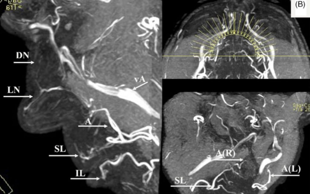
Results: A harmless visualisation of the facial arteries is currently only possible with US or MRA. US may identify single vessels but never the complete arterial network. A combination of IR “heat enhancement” and a MRA 3D-TOF sequence might make it feasible to visualize a large number of facial arteries in a risk-free, radiation-free, contrast-free and non-invasive way.
Conclusion: Currently, a new combination of IR “heat enhancement” and a MRA 3DTOF sequence might be the only method to visualize a large number of facial arteries.
Key takeaways:
- High-resolution ultrasonography is a non-invasive imaging technique that may even visualize small facial arteries, including depth measurements. However, it lacks the ability to visualize a wide area, show the 3D arterial vasculature, it is operator-dependent, and time-consuming. Additionally, combining US visualization with dermal filler injections simultaneously seems cumbersome.
- Conventional angiography remains the most precise evaluation technique with the highest sensitivity and specificity, but demands contrast medium, is an invasive procedure, and exposes the patient to harmful X-rays. It should therefore be avoided in aesthetic setting.
- CT angiography delivers a high-resolution and 3D visualization of the arterial anatomy. Although vessel catheterization is avoided, there is still a risk of complications due to the use of iodine contrast medium and the radiation exposure.
- 3D TOF MOTSA is a flow-based MRI technique that, in combination with facial heating, is able to visualize the 3D arterial anatomy of the face in a harmful way, without intravenous contrast, nor harmful X-rays. The main disadvantage is the risk of artefacts in case of metal objects in the face.